The Craniovertebral Junction Area and the Role of the Ligaments and Membranes

Neurosurgery 68:291–301, 2011 DOI: 10.1227/NEU.0b013e3182011262

Traumatic injuries of the craniovertebral junction (CVJ) area are common and frequently the outcome of motor vehicle accidents, falls, and diving accidents.

To define and characterize CVJ traumatic injuries, some international classifications are currently in use, and they are thought and focused on junction bone fracture. However, recent data point out a major important role of the CVJ ligaments and membranes in traumatic injuries with a secondary function of the osseous structures.

Emphasizing the correct role of the ligaments and membranes is extremely important for determining appropriate medical or surgical planning for patients and also to design new CVJ injury classifications.

We reviewed every recent major publication on the ligaments and membranes of the CVJ area. We divided the information into sections concerning anatomy, embryology, biomechanics, trauma, and CVJ bone fractures.

A role of the ligaments and membranes in the traumatic injuries of the CVJ area has often been recognized; but only recently, with the increase in the knowledge of the anatomic and biomechanical junction area, supported by neuroradiological tools (magnetic resonance imaging) and a more detailed traumatic injuries assessment, has the role of the ligaments and membranes been highlighted.

Ligaments and membranes have a pivotal role in each junctional ability and are the key to orienting any medical or surgical indications in this unique area of the spine.

Development of Stereotactic Mass Spectrometry for Brain Tumor Surgery

Neurosurgery 68:280–290, 2011 DOI: 10.1227/NEU.0b013e3181ff9cbb

Surgery remains the first and most important treatment modality for the majority of solid tumors. Across a range of brain tumor types and grades, postoperative residual tumor has a great impact on prognosis. The principal challenge and objective of neurosurgical intervention is therefore to maximize tumor resection while minimizing the potential for neurological deficit by preserving critical tissue.

OBJECTIVE: To introduce the integration of desorption electrospray ionization mass spectrometry into surgery for in vivo molecular tissue characterization and intraoperative definition of tumor boundaries without systemic injection of contrast agents.

METHODS: Using a frameless stereotactic sampling approach and by integrating a 3-dimensional navigation system with an ultrasonic surgical probe, we obtained image-registered surgical specimens. The samples were analyzed with ambient desorption/ ionization mass spectrometry and validated against standard histopathology. This new approach will enable neurosurgeons to detect tumor infiltration of the normal brain intraoperatively with mass spectrometry and to obtain spatially resolved molecular tissue characterization without any exogenous agent and with high sensitivity and specificity.

RESULTS: Proof of concept is presented in using mass spectrometry intraoperatively for real-time measurement of molecular structure and using that tissue characterization method to detect tumor boundaries. Multiple sampling sites within the tumor mass were defined for a patient with a recurrent left frontal oligodendroglioma, World Health Organization grade II with chromosome 1p/19q codeletion, and mass spectrometry data indicated a correlation between lipid constitution and tumor cell prevalence.

CONCLUSION: The mass spectrometry measurements reflect a complex molecular structure and are integrated with frameless stereotaxy and imaging, providing 3-dimensional molecular imaging without systemic injection of any agents, which can be implemented for surgical margins delineation of any organ and with a rapidity that allows real-time analysis.

Facet Joint Biomechanics at the Treated and Adjacent Levels After Total Disc Replacement

SPINE Volume 36, Number 1, pp E27–E32

Study Design. Biomechanical study using human cadaveric lumbar spines.

Objective. To evaluate effects of total disc replacement (TDR) on spine biomechanics at the treated and adjacent levels.

Summary of Background Data. Previous studies on spine biomechanics after TDR were focused on facet forces and range of motion and report contradictory results. Characterization of contact pressure, peak contact pressure, force, and peak force before and after TDR may lead to a better understanding of facet joint function and may aid in prediction of long-term outcomes after TDR.

Methods. Seven fresh-frozen human cadaveric lumbar spines were potted at T12 and L5 and installed in a 6 degrees of freedom displacement- controlled testing system. Displacements of 15° flexion/ extension, 10° right/left bending, and 10° right/left axial rotation were applied. Contact pressure, peak contact pressure, force, peak force, and contact area for each facet joint were recorded at L2–L3 and L3–L4 both before and after TDR at L3–L4. The data were analyzed with analysis of variance and t tests.

Results. Axial rotation had the most impact on contact pressure, peak contact pressure, force, peak force, and contact area in intact spines. During lateral bending and axial rotation, TDR resulted in a significant increase in facet forces at the level of treatment and a decrease in contact pressure, peak contact pressure, and peak force at the level superior to the TDR. With flexion/extension, there was a decrease in peak contact pressure and peak contact force at the superior level.

Conclusion. Our study demonstrates that rotation is the most demanding motion for the spine. We also found an increase in facet forces at the treated level after TDR. We are the first to show a decrease in several biomechanical parameters after TDR at the adjacent superior level. In general, our findings suggest there is an increase in loading of the facet joints at the level of disc implantation and an overall unloading effect at the level above.

Vascular Endothelial Growth Factor: The Major Factor for Tumor Neovascularization and Edema Formation in Meningioma Patients

Neurosurgery 67:1703–1708, 2010 DOI: 10.1227/NEU.0b013e3181fb801b

Peritumoral brain edema (PTBE) may be crucial in the clinical outcome of meningioma patients. The underlying pathogenetic key mechanism has so far not been determined. Sex, age, tumor size, location, involvement of other structures, or the histological appearance was not found to sufficiently explain PTBE formation in meningiomas.

OBJECTIVE: As PTBE formation is widely accepted to be vasogenic, we investigated the role of vascular endothelial growth factor (VEGF) and pial supplying vessels in a series of World Health Organization (WHO) grade I meningiomas.

METHODS: A total of 79 patients with WHO grade I meningiomas were immunohistochemically studied for VEGF and MIB-1. Pre- and postoperative magnetic resonance imaging including 3-dimensional reconstruction of 1.3-mm thick layers, with calculation of tumor and edema volume, was performed. Intraoperatively, the vascular supply and arachnoidal state were noted by the neurosurgeon.

RESULTS: VEGF was found to be exclusively confined to meningioma tumor cells. We identified 4 different patterns. VEGF and supplying pial vessels were found in 14 meningioma patients, pial vascular supply only in 3, VEGF expression only in 46, and neither VEGF expression nor supplying pial vessels in 16. Only the occurrence of both pial vascular supply and tumor VEGF expression was found to be correlated with PTBE formation (P , .002).

CONCLUSION: Our data suggest that VEGF may be crucial in angiogenesis and therefore indirectly in PTBE formation in World Health Organization grade I meningiomas

Minimally invasive endoscopic transventricular hemispherotomy for medically intractable epilepsy: a new approach and cadaveric demonstration

J Neurosurg Pediatrics 6:000–000, 2010. (DOI: 10.3171/2010.9.PEDS10267)

Surgery for medically intractable epilepsy secondary to unihemispheric pathology has evolved from more aggressive hemispherectomy to less aggressive variations of hemispherotomy. The authors propose a novel minimally invasive endoscopic hemispherotomy that should give results comparable to conventional open craniotomy and microsurgery.

Methods. Endoscopic transventricular hemispherotomy was performed in 5 silicon-injected cadaveric heads in the authors’ minimally invasive neurosurgery laboratory. The lateral ventricle was accessed endoscopically through a frontal and occipital bur hole. White matter disconnections were performed to unroof the temporal horn and to disconnect the frontobasal region, corpus callosum, and fornix.

Results. Using an endoscopic transventricular approach, all white matter disconnections were successfully performed in all 5 cadavers.

Conclusions. The authors have demonstrated the feasibility of endoscopic transventricular hemispherotomy in a cadaveric model. The technique is simple and could be useful in a subgroup of patients with parenchymal volume loss and ventriculomegaly.

Visualization of the Anterior Cerebral Artery Complex With a Continuously Variable-View Rigid Endoscope: New Options in Aneurysm Surgery

Neurosurgery 67[ONS Suppl 2]:ons321–ons324, 2010 DOI: 10.1227/NEU.0b013e3181f74548.

Neuroendoscopy is increasingly used as an adjunctive tool in intracranial aneurysm surgery.

OBJECTIVE: To assess the versatility of a prototype continuously variable-view rigid endoscope in visualizing the anterior cerebral artery complex.

METHODS: In 5 formaldehyde-fixed, arterially injected specimens, a standard frontolateral approach was used on both sides. After meticulous microsurgical dissection using this approach, the prototype of a multivariable rigid endoscope (EndoCAMeleon; Karl Storz GmbH & Co, Tuttlingen, Germany) was inserted. It is a rigid endoscope that is capable of changing its angle of view while remaining stationary and shape invariant. We inspected the anterior cerebral artery complex, using and testing the capabilities of the device.

RESULTS: The continuously variable viewing mechanism enables the surgeon to adjust the field of view continuously and to optimize the visualization of the neurovascular structures. Because of the rigid tip combined with the continuously variable viewing mechanism, the need to move the endoscope within the surgical field was minimal. The field of view changes, but the tip itself hardly moves. The EndoCAMeleon was able to enhance both the visibility of the anterior cerebral artery complex and the accessibility of the A1 and A2 arterial walls to a range of approximately 270 degrees.

CONCLUSION: The EndoCAMeleon enhances the visibility of the anterior cerebral artery complex and facilitates endoscope-assisted inspection, planning of clip application, and clip control.

Tractography of the amygdala and hippocampus: anatomical study and application to selective amygdalohippocampectomy

J Neurosurg 113:1135–1143, 2010.(DOI: 10.3171/2010.3.JNS091832)

The aim of this study was to evaluate, using diffusion tensor tractography, the white matter fibers crossing the hippocampus and the amygdala, and to perform a volumetric analysis and an anatomical study of the connections of these 2 structures. As a second step, the authors studied the white matter tracts crossing a virtual volume of resection corresponding to a selective amygdalo-hippocampectomy.

Methods. Twenty healthy right-handed individuals underwent 3-T MR imaging. Volumetric regions of interest were manually created to delineate the amygdala, the hippocampus, and the volume of resection. White matter fiber tracts were parcellated using the fiber assignment for continuous tracking tractography algorithm. All fibers were registered with the anatomical volumes.

Results. In all participants, the authors identified fibers following the hippocampus toward the fornix, the splenium of the corpus callosum, and the dorsal hippocampal commissure. With respect to the fibers crossing the amygdala, the authors identified the stria terminalis and the uncinate fasciculus. The virtual resection disrupted part of the fornix, fibers connecting the 2 hippocampi, and fibers joining the orbitofrontal cortex. The approach created a theoretical frontotemporal disconnection and also interrupted fibers joining the temporal pole and the occipital area.

Conclusions. This diffusion tensor tractography study allowed for good visualization of some of the connections of the amygdala and hippocampus. The authors observed that the virtual selective amygdalohippocampectomy disconnected a large number of fibers connecting frontal, temporal, and occipital areas

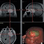
Skull base tumor model. Laboratory investigation

J Neurosurg 113:1106–1111, 2010. DOI: 10.3171/2010.3.JNS09513

Resident duty-hours restrictions have now been instituted in many countries worldwide. Shortened training times and increased public scrutiny of surgical competency have led to a move away from the traditional apprenticeship model of training. The development of educational models for brain anatomy is a fascinating innovation allowing neurosurgeons to train without the need to practice on real patients and it may be a solution to achieve competency within a shortened training period. The authors describe the use of Stratathane resin ST-504 polymer (SRSP), which is inserted at different intracranial locations to closely mimic meningiomas and other pathological entities of the skull base, in a cadaveric model, for use in neurosurgical training.

Methods. Silicone-injected and pressurized cadaveric heads were used for studying the SRSP model. The SRSP presents unique intrinsic metamorphic characteristics: liquid at first, it expands and foams when injected into the desired area of the brain, forming a solid tumorlike structure. The authors injected SRSP via different passages that did not influence routes used for the surgical approach for resection of the simulated lesion. For example, SRSP injection routes included endonasal transsphenoidal or transoral approaches if lesions were to be removed through standard skull base approach, or, alternatively, SRSP was injected via a cranial approach if the removal was planned to be via the transsphenoidal or transoral route. The model was set in place in 3 countries (US, Italy, and The Netherlands), and a pool of 13 physicians from 4 different institutions (all surgeons and surgeons in training) participated in evaluating it and provided feedback.

Results. All 13 evaluating physicians had overall positive impressions of the model. The overall score on 9 components evaluated—including comparison between the tumor model and real tumor cases, perioperative requirements, general impression, and applicability—was 88% (100% being the best possible achievable score where the evaluator strongly agreed with the proposed factor). Individual components had scores at or above 80% (except for 1). The only score that was below 80% was related to radiographic visibility of the model for adequate surgical planning (score of 74%). The highest score was given to usefulness in neurosurgical training (98%).

Conclusions. The skull base tumor model is an effective tool to provide more practice in preoperative planning and technical skills.